TISSUE ENGINEERING
Creating Tissues and Saving Lives

TIMELINE
1500s
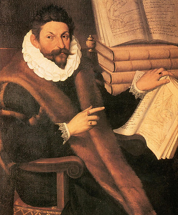
Gaspare Tagliacozzi was an Italian surgeon who worked with reconstructive surgery in Bologna. He improved on the method used to repair noses that had been amputated. He took skin from the arm (this is called a pedicle). However, the pedicle was still attached to the arm. The pedicle was attached to the nose, and the patient's arm was bandaged so the arm wouldn't move. After 20 days, the pedicle was severed from the arm. After another 14 days, the pedicle was shaped so that it looked like the nose.
(T.1)
(I.1)
1665
In 1665, Robert Hooke discovered cells. However, Hooke had been looking at the cell walls of cork tissue. He described discoveries in his book Micrographia. He also coined the word 'cells', as the cell walls he observed reminded him of the cells in a monastery.
(T.2)
The "cells" that Hooke observed under his microscope
(I.2)
1822
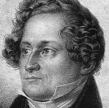
Johann Friedrich Dieffenbach worked with skin grafts. His experiments with animal and clinical transplantation are described in his work Nonnulla de Regeneratione et Transplantatione. He also described a way to use pedicled skin flaps (similar to Tagliacozzi). Dieffenbach is considered one of the modern founders of reconstructive surgery.
(T.3)
1838-1839
Theodor Schwann, Matthias Jakob Schleiden, and Rudolph Virchow created the first Cell Theory. The Cell Theory consists of three parts:
-
"All living organisms are made of cells"
-
"The cell is the basic unit of life"
-
"Cells come from pre-existing cells"
(T.4)

3-D rendering of cells under a microscope
1954
In 1954, the first successful kidney transplant was performed by Joesph E. Murray at Peter Bent Brigham Hospital in Boston. The kidney was transferred from one twin to another. Scientists predicted that the new kidney wouldn't be rejected by the recipient's body since the twins' immune systems are similar.
(T.5)

Joseph E. Murray performing the first successful kidney transplant
1970s
A pediatric orthopedic surgeon at the Children’s Hospital, W. T. Green, M.D., performed experiments where he tried to generate cartilage using chondrocytes on pieces of bone and implanting them in mice. Although he was unsuccessful, he showed that it would be possible to create tissue by planting cells on scaffolds, which is the basis of tissue engineering.
(T.6)

Diagram of a chondrocyte
1980s
In the 1980s, the term 'tissue engineering' was loosely applied to the surgical manipulation of tissues. However, it didn't mean what it meant today. The first recorded use of 'tissue engineering' as it is used today was in a published article titled “Functional Organ Replacement: The New Technology of Tissue Engineering” in Surgical Technology International in 1991.
(T.6)

Current issue of Surgical Technology International
1991
A young patient with Poland's Syndrome, a rare birth defect where there is an underdevelopment or absence of the pectoralis major, became the first person to receive a tissue-engineered implant. It was made from a synthetic polymer scaffold seed with autologous chondrocytes.
(T.6)

Diagram explaining Poland's Syndrome
1994
The Tissue Engineering Society, today is known as TERMIS, was founded by Drs. Charles A. and Joseph P. Vacanti in 1994.
(T.6)

1997
In 1997, Drs. Joseph and Charles Vacanti performed the infamous “mouse with human ear” experiment. They implanted the shape of a human ear on the back of a mouse in order to understand how scientists could grow body parts.
(T.7)

The "mouse with the human ear", or better known as the Vacanti mouse

2006
In 2006, Dr. Anthony Atala grew bladders for 7 children suffering from spina bifida, a birth defect that causes a baby's spinal cord to not develop properly. This marks the first time that artificial lab-grown organs have been implanted in humans.
(T.8)

Dr. Atala places a bladder-shaped mold seeded with bladder cells in a growth solution
2018
A new 3-D printing technique developed at the Imperial College in London can create structures that can mimic the mechanical properties of organs such as the brain and lung. This technique could lead to the creation of brain and lung tissues and eventually whole organs, which are incredibly hard to accomplish in regenerative medicine.
(T.9)

These images show the results of the experiment conducted by the Imperial College. A and B show the structure of the 3-D printed scaffold. C is when it is frozen, and D is the result.